
Horse Lameness Treatment:
A Complete Guide for Horse Owners
📋 Key Takeaways
Lameness is one of the most common - and costly - problems in performance horses. But it's also one of the most treatable when caught early.
- Most forelimb lameness starts in the foot. Around 90% of cases involve structures from the knee down.
- The right treatment depends entirely on an accurate diagnosis first. Treating the wrong thing doesn't work.
- Front and hind leg lameness have different causes, different treatment approaches, and different prognoses.
- Laminitis can look like a stone bruise at first. Waiting to call your vet often turns a manageable problem into a crisis.
- A multimodal approach - combining multiple therapies - produces the best outcomes for severe or complex cases.
You noticed your horse favoring one leg during warmup. Or maybe they've just felt "off" for a few days - not obviously lame, but not right either. Horse lameness treatment is one of the most searched topics in equine health, and for good reason.
Lameness is the most common cause of lost training days and early retirement in performance horses. But "lame" isn't a diagnosis - it's a symptom. The treatment depends entirely on what's causing it.
This guide walks through how vets find the cause of lameness, what treatment options exist by location and severity, when to call your vet immediately, and what a real-world recovery from a near-fatal case actually looks like.
What Causes Lameness in Horses?
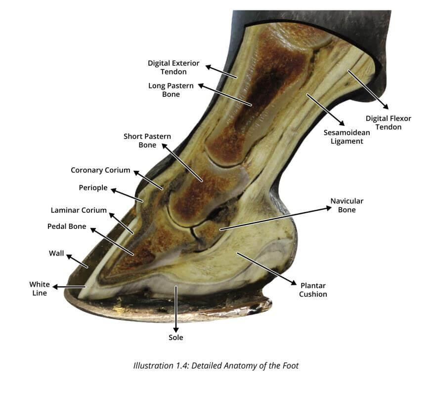
Understanding the cause of lameness in horses is the first step toward treating it. Most lameness is pain-driven - the horse changes its movement to protect a structure that hurts. Less commonly, the cause is neurological or mechanical.
In performance horses, causes fall into five main categories:
- Hoof and foot problems: Abscesses, bruising, navicular disease, white line disease, laminitis. Roughly 90% of front leg lameness starts somewhere below the knee.
- Joint problems: Arthritis in the hocks, stifles, fetlocks, or coffin joint. Very common in older performance horses and those with conformation that puts extra stress on certain joints.
- Tendon and ligament injuries: The suspensory ligament and the tendons running down the back of the lower leg are among the most commonly injured structures in performance horses. Overwork, bad footing, and sudden missteps are the usual causes.
- Back and hip pain: Often a knock-on effect of a problem lower in the leg. When a horse hurts somewhere in a limb, they compensate - and over time that puts strain on the back.
- Neurological causes: Less common, but conditions affecting the nervous system can produce gait changes that look a lot like lameness. Your vet will rule this in or out during the evaluation.
A horse can also have more than one thing going on at the same time. A horse with sore hocks may develop back pain from compensating. Treating one problem while missing another is one of the main reasons horses don't improve the way they should.
How Vets Diagnose Horse Lameness
Before any treatment can begin, a vet needs to find exactly where the pain is coming from. A thorough lameness evaluation is systematic and precise. Here's what it involves:
Step 1: Watching Your Horse Move
The vet watches your horse walk and trot - in a straight line, on a circle, in both directions, on hard ground and soft. They're looking for a head bob (which points to a front leg), a hip that drops more on one side than the other (which points to a hind leg), a shortened stride, or anything that looks different going one direction versus the other.
Step 2: Flexion Tests
The vet holds a leg in a bent position for about a minute, then has the horse trotted immediately. If the horse comes out noticeably more lame, that joint or the area around it is likely involved. It's a simple but reliable way to narrow things down.
Step 3: Nerve Blocks
This is where the detective work gets precise. The vet uses a small injection of local anaesthetic - the same type used at a dentist - to temporarily numb a specific area of the leg. If the horse trots sound after the block, the pain was coming from that area. The vet works from the foot upward, one region at a time, until the source is pinpointed.
Step 4: Imaging
Once the area is confirmed, imaging shows exactly what's going on inside:
- X-rays - bones and joints
- Ultrasound - tendons, ligaments, and soft tissue
- MRI - highly detailed view of the foot, especially useful for navicular disease
- CT scan - used for complex bone injuries
- Bone scan - picks up areas of active bone change when other imaging hasn't given a clear answer
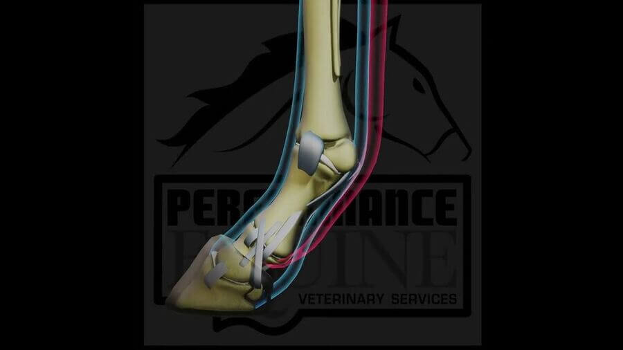
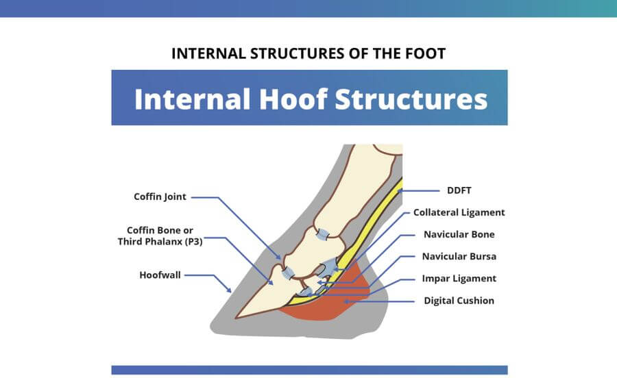
Check out Dr. Rullan's free guide on the anatomy and radiographs of a horse's foot to learn more.
Lameness Grading Scale (AAEP 0–5)
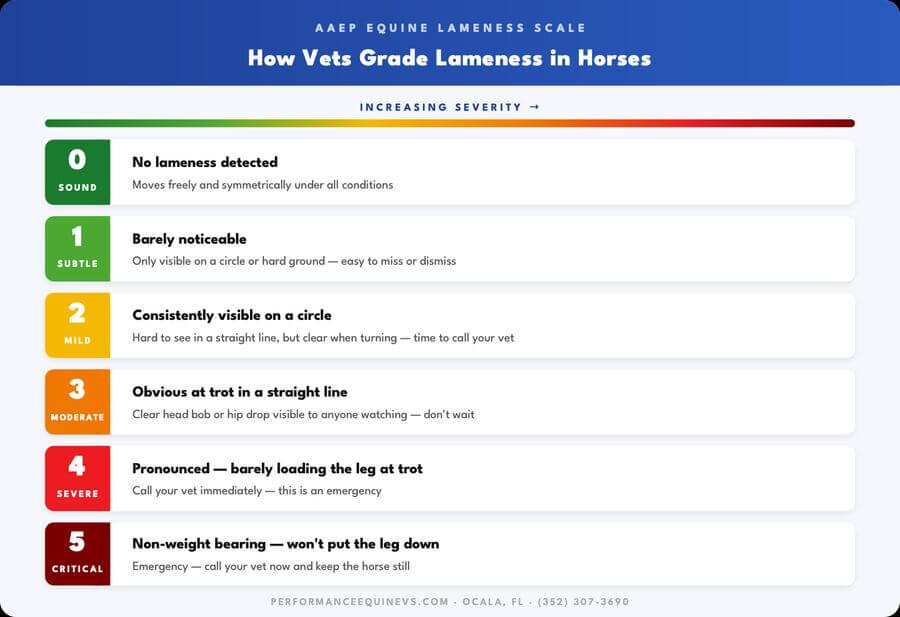
Vets use this standardized scale to describe severity. The grade guides treatment decisions and helps track progress over time.
Grades 1–2 often correspond to early or developing problems - the most valuable time to intervene. Grades 4–5 are emergencies.
Horse Lameness Treatment by Location
The most effective horse lameness treatment depends on where the problem is, what structure is involved, and how severe it is.
Front Leg Lameness Treatment
Front leg lameness in horses accounts for the majority of performance horse cases. The foot and lower limb are most commonly involved.
- Hoof abscess: Soaking, poulticing, and draining the abscess. Most horses are back to normal within a few days to two weeks.
- Navicular disease: Special shoeing to change how the foot lands and loads, anti-inflammatory medication, and injections into the foot to reduce pain and inflammation. In advanced cases, MRI helps guide a more targeted approach.
- Laminitis: Treated as an emergency. Deep shavings or sand bedding, special shoeing to take pressure off the rotating bone, pain management, and strict diet control to address the underlying cause. See our full guide to laminitis treatment.
- Tendon and ligament injuries: Controlled rest, cold therapy, and a carefully structured return to work. Regenerative treatments like PRP (where healing factors from the horse's own blood are injected into the injury) or stem cell therapy can significantly improve the quality of the tissue that heals. Water treadmill rehab helps rebuild strength without putting too much strain on the healing structure. Dr. Rullan's guide on recovering from tendon injuries covers more on this.
- Arthritis in the lower leg joints: Injections to reduce inflammation and lubricate the joint. Shockwave therapy for longer-standing cases. The goal is managing the condition so the horse stays comfortable and functional.
Hind Leg Lameness Treatment
Hind leg lameness in horses is often subtler than front limb lameness and easier to miss. The hocks and stifles are the most common sites.
- Hock arthritis: One of the most common causes of hind leg lameness. Hock injections to reduce inflammation and lubricate the joint work well for most horses. In cases where the lower hock joints are the problem, they can sometimes be fused surgically - which sounds dramatic but often results in a pain-free, fully functional horse.
- Stifle problems: A stifle that locks up, bone or cartilage fragments, or soft tissue injuries. Treatment ranges from exercise adjustments and injections to keyhole surgery to remove fragments if they're causing ongoing problems.
- Hind suspensory ligament injury: Rest, regenerative therapy, and a very gradual return to work. This injury tends to be more serious than the same injury in the front leg and needs to be managed carefully to avoid re-injury.
- Lower back and hip joint pain: Injections guided by ultrasound to precisely target the right area, combined with rehabilitation and bodywork. Often a sign that something else is off in a hind leg - which is why the whole picture needs to be assessed.
Hind leg lameness can be trickier to spot and, depending on the structure involved, harder to get back to full performance. That makes getting on it early even more important.
Treatment Options at a Glance
Most cases require more than one approach. Here's a quick reference to the main treatment modalities, what they're used for, and what to know about each.
| Treatment | Best Used For | Key Notes |
|---|---|---|
| Rest + controlled exercise | All tendon and ligament injuries | Unstructured rest alone rarely works - how you bring the horse back matters as much as the rest itself |
| Pain relief (bute, Equioxx) | Acute pain and inflammation | Manages discomfort while the real treatment takes effect - doesn't fix the underlying problem |
| Joint injections | Arthritis, joint inflammation | Anti-inflammatory medication injected directly into the joint. Very effective; how often depends on the horse and the condition |
| Therapeutic shoeing | Foot and tendon conditions | Special shoeing changes how the foot loads and moves. Essential for laminitis, navicular disease, and most chronic foot problems |
| Shockwave therapy | Tendon, ligament, and bone injuries | Uses sound waves to stimulate healing. Non-invasive and effective for longer-standing soft tissue problems |
| PRP | Tendon and ligament injuries | Healing factors concentrated from the horse's own blood and injected directly into the injury to speed and improve healing |
| Stem cell therapy | Tendon, ligament, cartilage | Encourages the body to build better-quality tissue at the injury site. Often used alongside PRP |
| Hyperbaric oxygen therapy | Laminitis, wounds, post-surgery | Pressurised oxygen increases the amount available to healing tissue by up to 600%. Particularly useful where blood supply is compromised |
| Water treadmill | Rehab and conditioning | Lets the horse exercise and build muscle without putting full weight through a healing structure |
| Surgery | Bone fragments, fractures, severe joint disease | Only when conservative options have been tried or clearly won't work |
Real-World Recovery: Stewart's Story
Sometimes the best way to understand what's possible with horse lameness treatment is to look at a case that wasn't supposed to make it.
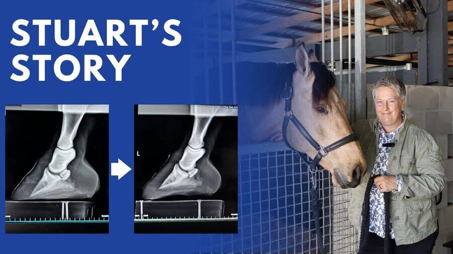
Patient: Stewart | 8-year-old Hanoverian warmblood gelding
Owner: Dr. Bess Darrow, equine veterinarian and dentist, Ocala, FL
Diagnosis: Catastrophic bilateral laminitis with coffin bone rotation and sinking
Initial prognosis given: Euthanasia
Outcome: Sound and back under saddle exactly one year later
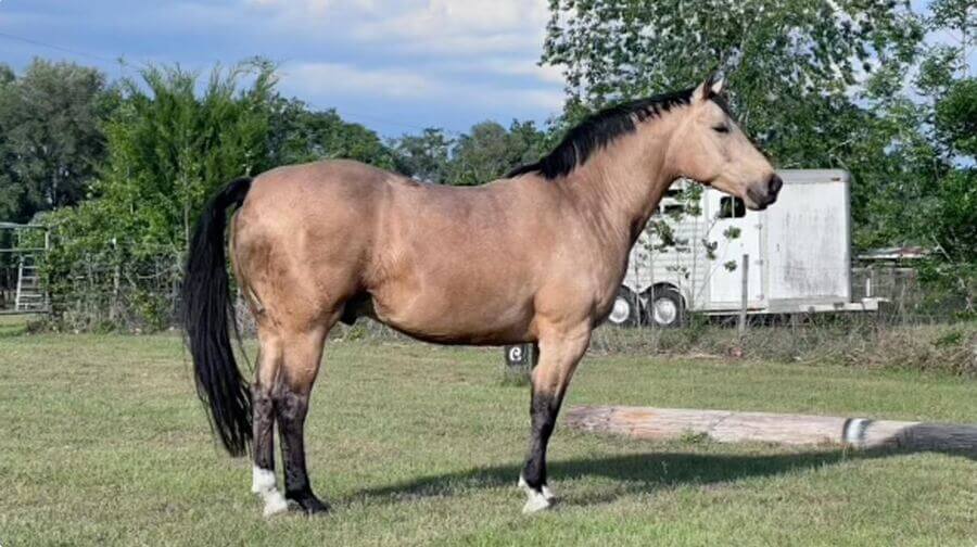
Stewart was an 8-year-old Hanoverian warmblood owned and raised by Dr. Bess Darrow, an equine veterinarian in Ocala. Despite receiving expert care from birth, Stewart developed insulin resistance around age seven - gaining weight steadily and showing subtle lameness that Dr. Darrow initially attributed to the hard, drought-affected ground in her arena.
Then one morning she found him in excruciating bilateral lameness on both front feet. A colleague took radiographs. The diagnosis was catastrophic laminitis - coffin bone rotation and sinking through the soles of both hooves. The recommendation was euthanasia.
The radiographs below show exactly what that looked like - and what the foot looked like a year later.
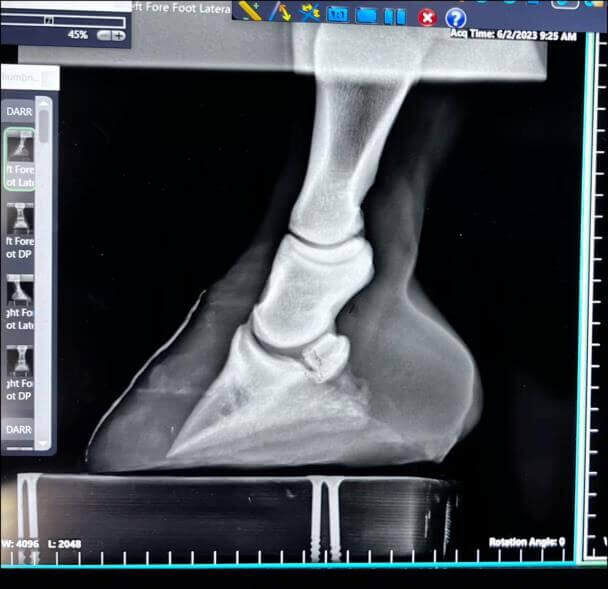
Severe coffin bone rotation and sinking
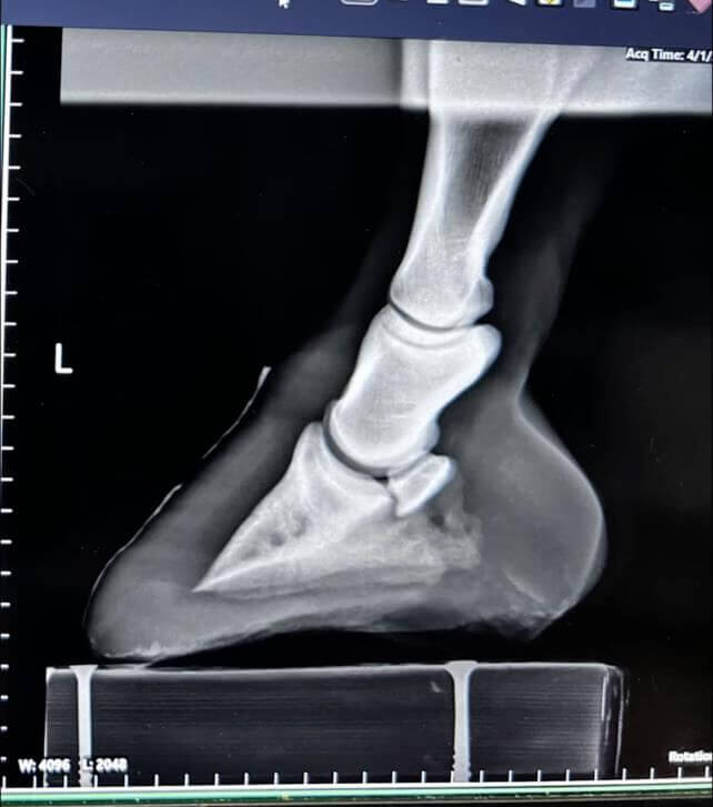
Coffin bone returned to correct anatomical alignment
Dr. Darrow had been undergoing hyperbaric oxygen treatments herself for a separate condition and had seen how they supported healing in tissues with compromised circulation - exactly the mechanism at play in laminitis. Rather than accepting the prognosis, she brought Stewart to EPIC.
The Treatment Protocol
Dr. Rullan and the EPIC team implemented what he describes as a multimodal approach - multiple therapies working together, not in isolation:
- Hyperbaric oxygen therapy: 5 days a week for 30 days. Increases oxygen availability to damaged tissue by up to 600% and mobilizes stem cells from bone marrow to support healing.
- Therapeutic shoeing: Correcting the mechanics of the foot, supporting the coffin bone position, and promoting healthy new hoof growth.
- Diet management: Strict low-sugar, low-starch feeding to control the underlying insulin resistance and reduce load on the damaged feet.
- Holistic support: Acupuncture, chiropractic work, and massage to address secondary strain on the hindquarters and back - which take on enormous load when the front feet are too painful to bear weight.
- Mental stimulation: Daily enrichment - clicker training, ball games, interaction with staff - to prevent the depression that commonly accompanies months of stall confinement.
After 30 days of hyperbaric therapy, Stewart walked out of EPIC in hoof boots and loaded onto a trailer to go home. That was just the beginning. Over the following year, he grew an entirely new hoof capsule. His coffin bones returned to correct anatomical alignment. Exactly one year after his diagnosis, he went back into regular ridden work.
What Made the Difference
Dr. Rullan is direct about what separated Stewart's outcome from cases that don't go as well:
- Early detection of the insulin resistance, even though the laminitis still became severe
- Aggressive, multimodal treatment rather than a wait-and-see approach
- An owner with the knowledge and commitment to continue the full protocol at home after discharge
- A team that missed nothing - no treatment was skipped, delayed, or deprioritized
When to Call Your Vet
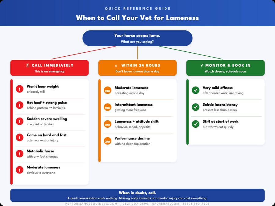
Not every lameness is an emergency. But some are - and waiting 24 hours on the wrong one can change the outcome significantly.
Call immediately
- Your horse won't put weight on a leg, or barely will
- The hoof feels hot and you can feel a strong, pounding pulse behind the pastern - this can mean laminitis
- There's sudden severe swelling in a joint or tendon
- The lameness came on hard and fast after a workout or a known injury
- Your horse has metabolic disease (insulin resistance, Cushing's) and is showing any foot-related changes
Call within 24 hours
- Moderate lameness that's been there for more than a day
- Intermittent lameness that's becoming more frequent
- Lameness alongside changes in attitude, appetite, or behavior
Keep an eye on it, get it booked in
- Very mild lameness after harder-than-usual work that seems to be getting better
- A subtle "offness" that comes and goes but has been there for more than a week
Frequently Asked Questions
How long does horse lameness treatment take?
It completely depends on what's causing it. A hoof abscess can clear up in a few days. A tendon injury can take anywhere from 6 to 18 months of careful rehabilitation. Arthritis is usually managed long-term rather than cured. The key is knowing exactly what you're dealing with and having a realistic plan from the start.
Can a lame horse recover completely?
Many do - but it depends on the injury. A horse with an abscess goes straight back to full work. A horse with a serious tendon injury may return to light work but not high-level competition. Stewart recovered from what should have been a fatal case of laminitis and went back under saddle a year later. What's possible is always worth discussing with your vet once you have a real diagnosis in hand.
Is laminitis always obvious?
No - and that's what makes it dangerous. Early laminitis can look like a mild stone bruise or a horse that's just a little off. By the time it's obviously lame, serious damage is often already done. Any horse with metabolic issues - easy keepers, horses with cresty necks or fat deposits - should be watched closely for any changes in how they move or feel in their feet.
What's the difference between bute and Equioxx?
Both are anti-inflammatory pain relievers. Bute (phenylbutazone) works quickly and is used for short-term pain management. Equioxx (firocoxib) is easier on the stomach and can be used for longer periods. Neither one fixes the underlying problem - they keep the horse comfortable while the treatment does its job.
Does my horse need surgery?
Probably not. Most lame horses get better with rest, shoeing changes, injections, and rehabilitation. Surgery comes up for specific things - removing bone or cartilage fragments, certain fractures, or when everything else has been tried and hasn't worked. A thorough workup will tell you whether it's even on the table.
Can lameness be prevented?
Not completely - but you can stack the odds in your favour. Regular farrier visits, good footing, conditioning that's appropriate for what you're asking the horse to do, annual soundness checks, and acting quickly when something seems off all make a real difference over a horse's career.
Final Thoughts
Effective horse lameness treatment always starts in the same place: knowing exactly what's causing it. The right approach for a horse with navicular disease looks completely different from the right approach for a tendon tear. Getting that answer right, quickly, is what determines everything that follows.
Stewart's story is an extreme example. But the lesson applies to every lame horse: don't wait, don't guess, and don't underestimate what's possible with the right team and the right approach.
If your horse is showing signs of lameness, schedule a lameness evaluation with the PEVS team or call us at (352) 307-3690. We're available Monday–Friday 8am–5pm, Saturday 8am–12pm, and for emergencies 24/7.
This article is for educational purposes only and is not a substitute for professional veterinary advice. If you have concerns about your horse's health or soundness, consult a licensed veterinarian.